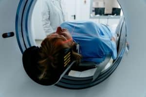
Do you know the difference between a CT Scan and an MRI? Continue to read this article to know the particularities of each. A computed tomography (CT) scan, or computerized axial tomography (CAT) scan, involves a patient lying down in a large X-ray machine known as the CT scanner. The CT scan uses X-rays to produce images. The scan uses ionizing radiation which has the potential to be damaging to tissues, but the amount used is very small to minimize this risk. As such, physicians consider CT scans to be safe despite the minimal radiation used.
How do CT Scans Work?
A CT scan only takes about 5 to 15 minutes, depending on the organ being studied. A contrast media may be given depending on what the scan is being performed for and the body part being imaged. This media can be administered orally (by mouth) and/or intravenously (through a needle).
The CT scanner will take multiple images of the body at once. These images will then be stacked together to form a 3D image of the body.

What is an MRI Scan?
Magnetic resonance imaging, or MRI, is a type of scan that uses radio waves and magnets to produce images. This makes MRI scans different from CT scans because it does not use ionizing radiation. MRI scan requires the use of a strong magnetic field and as such there are contraindications; this includes pacemakers, cochlear implants, and possibly bullets and any other foreign metals in the body depending on their location. Also, it can produce detailed images and is great for imaging the body.
How do MRI Scans Work?
The MRI scans work producing a strong magnetic field. The body has natural magnetic properties due to the abundance of hydrogen in water and fats within the body. When the body is in the MRI scanner, the hydrogen ions in the body all orient themselves in the same direction. This magnetic field can be adjusted depending on the body part being studied. When radio waves are added to the magnetic field, the hydrogen ions change direction. When the radio waves are removed, the hydrogen ions return to their resting state. This causes a signal to be emitted, thus creating the images we see.
For certain MRI studies, gadolinium-based contrast media may be given intravenously to improve diagnostic capability.
Both CT and MRI scan the body to produce internal images. MRI scans may have more detailed images, but both scans have important use and are helpful in screening for diseases. Your doctor will make sure that you receive the scan that is right for you.


